Difference between gestational sac and yolk sac?
 Gestational Sac vs Yolk Sac
Introduction:
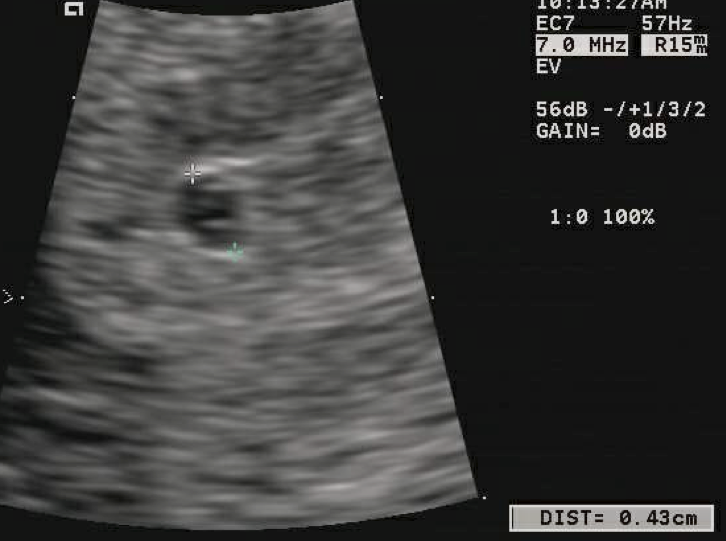
Gestational sac forms the earliest visible structure once fertilization takes place. It can be viewed on the ultrasonography on the upper wall of the uterus or the womb as a small hyper-echoic (dark) shadow surrounded by a hypo-echoic (light) shadow. The yolk sac is present in the gestational sac and is attached to the growing embryo on the front. The yolk sac is the nutritional source of the embryo in the early stages before the development of the circulation. The yolk sac provides nutrients in the circulation when the blood passes through it and later on returns back to embryo.
Difference in functions
Gestational sac is seen and used to confirm a positive intrauterine pregnancy even before the embryo can be viewed. Once the embryo develops as a small tissue, there is formation of yolk sac. The mean gestational sac diameter is an estimate for the gestational age. If the gestational sac reaches 20mm in length there should be appearance of yolk sac and when the gestational sac reaches 25mm there should be appearance of fetal pole in the embryo. The doctors thus can approximate the events on the basis of the gestational sac development.
Yolk sac functions as the developmental circulatory system in the tiny embryo before the actual internal circulation is formed. It the first element which can be seen at 3 days of gestation and it helps identify a true gestational sac. Yolk sac is covered by an external layer called the extra embryonic endoderm and outside which there is presence of one more layer called as extra embryonic mesoderm. The blood supply to the wall of the sac is by the primitive aorta and after the blood passes through a series of capillary mesh, returns to the tubular heart in the embryo by vitelline vessels. This is known as the vitelline circulation through which the embryo is provided with adequate nutrition.
By the end of the fourth week of gestation the yolk sac becomes a vesicle commonly known as the umbilical vesicle as the heart tube take over the function of the circulation in the embryo. The vesicle then is attached to the embryo by vitelline duct which gets obliterated by 7th week of gestation. If at all the vitelline duct does not obliterate then it will be seen as Meckel’s Diverticulum.
Gestational sac is visible at 5th week of gestation when actually the fetus is just 3 weeks by age. The gestational sac should grow 1mm per day after it is been detected by 5th week. This helps to identify if there is any abnormal growth in the pregnancy or if the gestational sac is growing normally.
Summary:
The gestational sac is a bigger sac which is formed as soon as fertilization takes place and is the first to be identified on ultra-sonography. The yolk sac is the nutritive sac formed within the gestational sac along with the embryo which will develop inside the gestational sac. The attachment of the various structures in the gestational sac like yolk sac, embryo as well as amniotic fluid will help verify the age of the fetus.
- Difference between near sightedness and far sightedness - January 21, 2015
- Difference between Diverticulosis and Diverticulitis - January 20, 2015
- Difference between Prilosec and Nexium - January 19, 2015
Search DifferenceBetween.net :
4 Comments
Leave a Response
References :
[0]http://commons.wikimedia.org/wiki/File:4_mm_gestational_sac.png

I think dark masses and cavities are hypoechoic rather hyperechoic as you have written regarding gestational sac. Please clear it up as it has confused me a lot. Thank you
Yes , you are very right. Am thinking am the only one seeing it.
It should be a mistake by the writer.
Thank you. Very helpful information.
Good one thanks