Difference Between Bullous Impetigo and Bullous Pemphigoid
Bullous impetigo is a condition of children where bullae form on the skin. Bullous pemphigoid is a skin condition of older adults where blisters form under the outer skin layer.

What is Bullous impetigo?
Definition:
A skin condition in which fluid-filled sacs (blisters) form on the top parts of the skin because of bacteria.
Causes and prevalence:
Bullous impetigo is caused by infection of the skin by bacteria. The bacterium causing the condition is Staphylococcus aureus. This is a fairly common condition that is found in about 10% of skin problems diagnosed in children.
Symptoms:
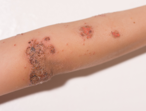
Vesicles form on the skin and become filled with fluid. These bullae then burst open and develop a golden-colored crust.
Complications:
A possible complication of bullous impetigo is cellulitis. This is when the skin becomes infected down into the deep layers of tissue.
Diagnosis:
Diagnosis of bullous impetigo is by the physical signs, the appearance of the lesions in a young child or infant. It is only if the treatment fails that samples may be taken to check the bacterial identification. This may be necessary if MRSA is suspected.
Treatment:
Treatment includes carefully washing the skin with soap and water and then applying an ointment. The ointments usually used to treat bullous impetigo include retapamulin or mupirocin antibiotic. The treatment can last from 5 to 7 days depending on what type of ointment is used.

What is Bullous pemphigoid?
Definition:
A condition where there is the formation of blisters underneath the epidermal layer of the skin.
Causes and prevalence:
The causes of bullous pemphigoid include viruses (specifically herpes viruses), radiation therapy and some drugs. It is quite a rare condition which seems to be an autoimmune response; it affects only about 23 people per million.
Symptoms:
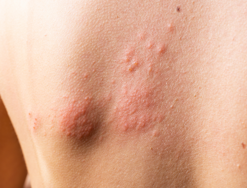
Signs of bullous pemphigoid include pruritis (itching) and the formation of bullae that are blisters that are full of fluid. These blisters are often red in color.
Complications:
Complications of the condition include bacterial skin infections and sepsis. Sepsis can lead to shock and death.
Diagnosis:
A skin biopsy combined with microscopy and specific enzyme-linked immunosorbent assays (ELISAs) can diagnose bullous pemphigoid. The ELISA helps by showing if specific autoantibodies are present in the skin. Certain of these autoantibodies can indicate if the patient has bullous pemphigoid.
Treatment:
The condition is treated with corticosteroids like prednisone, and anti-inflammatory drugs. The anti-inflammatory drugs are helpful if the skin is very inflamed. Immunosuppressants are also sometimes prescribed to help treat bullous pemphigoid. The immunosuppressants can be helpful since bullous pemphigoid has an autoimmune component.
Difference between Bullous impetigo and Bullous pemphigoid?
Definition
Bullous impetigo is a skin condition where fluid-filled blisters develop. Bullous pemphigoid is a skin condition where blisters form underneath the top layer of skin.
Layer of skin affected
In the case of bullous impetigo, it is the superficial, top layer of skin that is affected and where blisters form. In the case of bullous pemphigoid, it is the lower layers beneath the epidermis that is affected and is where blisters are formed.
Blisters
The blisters of bullous impetigo are fluid-filled, burst open easily, and develop a distinctive golden-colored crust. The blisters of bullous pemphigoid are subepidermal and look like red blotches.
Location of rash
The rash of bullous impetigo is often on the arms, legs, and torso. The rash of bullous pemphigoid is often on the armpits, thighs, and belly.
Age of patient
Children who are younger than 2 years tend to get bullous impetigo. Older adults of 60 to 80 tend to get bullous pemphigoid.
Diagnosis
Bullous impetigo is diagnosed by the physical symptoms. Bullous pemphigoid is diagnosed by microscopy and ELISA where autoantibodies are detected.
Treatment
The treatment of bullous impetigo includes using retapamulin or mupirocin antibiotic and making sure the skin is washed properly with soap and water. Anti-inflammatory and immunosuppressant medicines, and corticosteroid medicines are used to treat bullous pemphigoid.
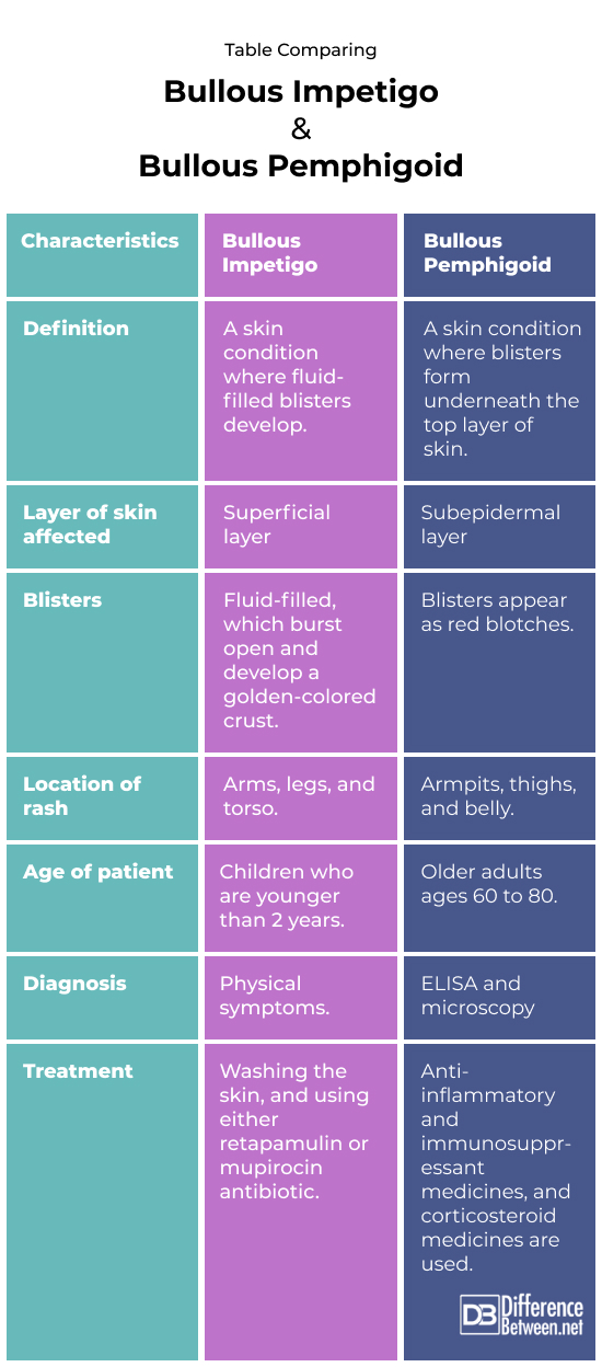
Table comparing Bullous impetigo and Bullous pemphigoid

Summary of Bullous impetigo Vs. Bullous pemphigoid
- Bullous impetigo is a skin condition of children while bullous pemphigoid occurs in older adults.
- The blisters of bullous impetigo develop a golden crust.
- The blisters of bullous pemphigoid appear as red blotches on the skin.
- Bullous impetigo and bullous pemphigoid need to be treated to reduce the risk of complications.
FAQ
Is bullous impetigo autoimmune?
No, bullous impetigo is an infectious disease not an autoimmune problem.
What do bullous pemphigoid blisters look like?
These are large blisters that occur often where there are skin folds or creases. The blisters are large, fluid-filled, and form red blotches on the skin.
What is the difference between bullous pemphigoid and pemphigus vulgaris?
Pemphigoid skin conditions affect the lower skin layers while pemphigus skin conditions affect the uppermost layers of the skin. Pemphigus vulgaris is also a condition where mouth ulcers commonly occur while these are rare in bullous pemphigoid.
How do you rule out pemphigoid?
A skin biopsy can be done to determine the cause of skin blisters and to exclude pemphigoid.
What is the confirmatory test for bullous pemphigoid?
A direct immunofluorescence (IF) test is done on a skin sample taken from around a blister. This is examined under a microscope and will show fluorescence if the condition is bullous pemphigoid.
- Difference Between Rumination and Regurgitation - June 13, 2024
- Difference Between Pyelectasis and Hydronephrosis - June 4, 2024
- Difference Between Cellulitis and Erysipelas - June 1, 2024
Search DifferenceBetween.net :
Leave a Response
References :
[0]Moro, Francesco, et al. "Bullous pemphigoid: trigger and predisposing factors." Biomolecules 10.10 (2020): 1432.
[1]Peraza, Daniel M. “Bullous Pemphigoid”. Merckmanuals. Merck & Co., 2022, https://www.msdmanuals.com/professional/dermatologic-disorders/bullous-diseases/bullous-pemphigoid
[2]Remus, Wingfield E. “Impetigo and Ecythema”. Merckmanuals. Merck & Co., 2022, https://www.msdmanuals.com/professional/dermatologic-disorders/bacterial-skin-infections/impetigo-and-ecthyma
[3]Image credit: https://www.canva.com/photos/MAD7oilF7mc-child-s-arm-with-impetigo/
[4]Image credit: https://www.canva.com/photos/MAE1zZJxwNw-skin-rash-and-blisters-on-body/
