Difference Between Centrosome and Centriole
The centrosome is an organelle that consists of two centrioles surrounded by a mass of proteins. The centriole is a structure that is made of microtubules that are arranged in a particular way.

What is Centrosome?
Definition of Centrosome:
The centrosome is a structure, an organelle that is found in the eukaryotic cell and is made of two centrioles surrounded by various proteins.
Size and structure of Centrosome:
The size of a centrosome is roughly double that of a centriole, since it consists of two centrioles. It also does not stay a constant size because it changes somewhat during the cell division process. The boundaries are really determined by the material that surrounds and encloses the centrioles rather than the centrioles themselves. The proteins that encircle the centrioles are said to form the pericentriolar material (PCM). The microtubules of the centrioles are able to attach to the proteins, and they are arranged at ninety-degree angles to each other.
Centrosome Formation:
In most species of mammals, the centrosome is believed to not be inherited from the parental cells, but rather to be formed anew in the cells of the zygote. The pericentriolar material is believed to help in producing the proteins and assembling the centrioles to form the centrosome.
Centrosome Location:
The centrosomes occur in animal cells and are typically found near to the nucleus in animal cells. It is copied during mitosis so that two centrosomes can be found on opposite sides of the nuclear envelope.
Function of Centrosome:
The centrosome is copied when a eukaryotic cell divides and has the function of forming the spindle which produces fibers that chromosomes attach to. The organelle plays a role in both the interphase and mitotic phases of the cell cycle. These organelles are important in organizing the microtubules, and in cell polarity. It is also involved in intracellular transport by organizing the microtubule array.

What is Centriole?
Definition of Centriole:
The centriole is a structure that is comprised of microtubules that are arranged in a particular manner. The microtubules are actually proteins that form short cylindrical structures.
Size and structure:
The microtubules form a centriole that has a length of about 200 nm wide and 500 nm long. A centriole is comprised of 9 microtubule sets. Each set is a triplet, in other words, it consists of 3 microtubules each. Research has shown that the microtubules arrange to form a cartwheel structure with proteins emanating out from a central component. These proteins connect to the A microtubule of each triplet. The other two microtubules are, unlike the A tubule, not complete and do not attach to the central part but attach to the A microtubule. The C tubule is the shortest of the three microtubules so at some parts there is a doublet rather than triplet structure present.
Formation of Centriole:
The protein SAS-6 has been found to be a molecule that acts as a precursor for the formation of centriole microtubules. In addition, scientists have found more protein molecules that seem to play a role in generating microtubules. Studies on C. elegans have shown that the protein SPD-2 organizes first, and then recruits more proteins including SAS-4, SAS-5, and SAS-6. These molecules come together and organize to produce the microtubular structure seen in a centriole.
Centriole Location:
Centrioles occur in some protist cells and in animal cells. They occur in pairs forming a centrosome but are also found at the bottom of cilia and flagella, where they occur as a single structure. In the case of flagella and cilia, the centrioles are found near the surface just inside the cell membrane where they are also referred to as basal bodies.
Function:
Where pairs of centrioles form a centrosome, they help form the spindle fibers during the process of cellular division. In the case where centrioles form the basal body of a cilium or flagellum, they help to actually produce the proteins that form the structure.
Difference between Centrosome and Centriole?
-
Definition
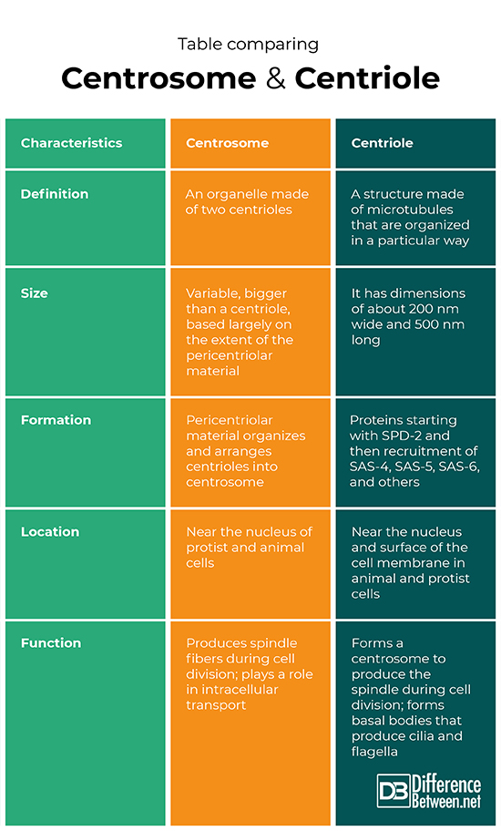
A centrosome is an organelle found in cells that consists of two centrioles. A centriole is a structure found in a cell that is comprised of microtubules that are arranged in a particular way.
-
Size
A centrosome is of variable size but always bigger than a centriole. A centriole has dimensions that are approximately 500 nm long and 200 nm wide.
-
Formation
The pericentriolar material helps form the centrosome by organizing the centrioles. Proteins starting with SPD-2 recruit other proteins such as SAS-4, SAS-5, and SAS-6 to form the centriole.
-
Location
The centrosome occurs near the nucleus, and after it has copied itself, on opposite sides of the nucleus. The centriole can occur either near the nucleus or near the cell membrane.
-
Function
The function of the centrosome is to produce the spindle during mitosis and to help regulate intracellular transport. The function of the centriole is to form the centrosome and to form the basal body that gives rise to cilia and flagella.
Table comparing Centrosome and Centriole

Summary of Centrosome Vs. Centriole
- A centrosome is an organelle that consists of two centrioles.
- A centriole is a structure made of microtubule proteins arranged in a particular way.
- A centriole is always smaller than a centrosome and also forms flagella and cilia.
- Both centrosomes and centrioles are found in animal cells and some protists.
- Difference Between Rumination and Regurgitation - June 13, 2024
- Difference Between Pyelectasis and Hydronephrosis - June 4, 2024
- Difference Between Cellulitis and Erysipelas - June 1, 2024
Search DifferenceBetween.net :
Leave a Response
References :
[0]Alieva, Irina B., and Rustem E. Uzbekov. "Where are the limits of the centrosome?" Bioarchitecture 6.3 (2016): 47-52.
[1]Azimzadeh, Juliette, and Wallace F. Marshall. "Building the centriole." Current biology 20.18 (2010): R816-R825.
[2]Ford, Jodie, Phillip Stansfeld, and Ioannis Vakonakis. "Coupling Form and Function: How the Oligomerisation Symmetry of the SAS-6 Protein Contributes to the Architecture of Centriole Organelles." Symmetry 9.5 (2017): 74.
[3]Image credit: https://en.wikipedia.org/wiki/File:Centrosome_Cycle.svg
[4]Image credit: https://upload.wikimedia.org/wikipedia/commons/thumb/6/6f/Centriole-en.svg/500px-Centriole-en.svg.png
