Difference Between Astrocyte and Microglia
The central nervous system (CNS), which includes the brain and spinal cord, is perhaps the most fascinating and essential of all organs in body. Two critically important neuroglial cell types are astrocytes and microglia. Their function is to communicate with neurons and provide biochemical support to the CNS.

What are astrocytes?
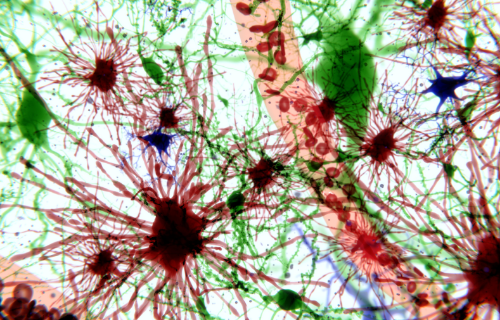
Astrocytes are macroglia cells and the most prevalent cell type in the CNS, making up anything between 20-50% of the volume of glia.
Astrocytes are highly active. In humans, a single astrocyte cell can interact with up to 2 million synapses at a time. Astrocytes play a number of roles in the CNS. These include
- generating proteins as well as cytokines, which signal proteins involved in the immune system,
- secreting and absorbing neural transmitters,
- providing nutrients to the nervous tissue,
- maintaining extracellular ion balance,
- repairing the brain and spinal cord following infection and traumatic injury, and
- maintaining the blood–brain barrier through providing biochemical support to the cells that form the blood–brain barrier.
Astrocytes regulate the transmission of electrical impulses and potassium within the brain, store and release glucose, and according to N. Swaminathan, regulate blood flow in the brain.
Astrocytes are important participants in various neurodevelopmental disorders. A. J. Barker and Ullian, for example, link astrocyte dysfunction to improper neural circuitry, and by virtue of that psychiatric disorders such as autism spectrum disorders and schizophrenia.

What are microglia?
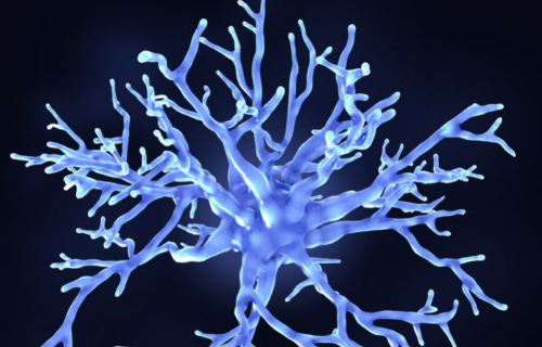
Microglia are a second type of neuroglial cell in the brain and spinal cord. Microglia inhabit about 5-20% of the mammalian brain and constantly move around within the CNS, engaging in extracellular signaling and identifying damaged neurons and infectious agents to maintain homeostasis. G. Kreutzberg suggests they are the first line of defense in brain pathologies, acting like macrophages or white blood cells that engulf and digest damaged or infected cells. The role of microglia is thus primarily defensive.
Microglial cells are extremely plastic; they adopt a specific form in response to the local conditions and the chemical signals they detect, and they undergo a variety of structural changes based on location and system needs. Microglial are also extremely sensitive to even small pathological changes in the central nervous system, reacting quickly to decrease inflammation and destroying infectious agents before they damage sensitive neural tissue after infectious agents have passed through the blood brain barrier.
Microglial cells immune response activity is a double-edged sword, however. Specifically, after becoming activated, microglia may release chemicals into the extracellular space to activate more microglia and start a cytokine storm. In other words, in addition to being able to destroy infectious organisms by engulfing foreign or damaged matter, microglia can also release a variety of cytotoxic substances in the process, such as hydrogen peroxide, nitric oxide, glutamate, aspartate, and quinolinic acid. These chemicals can damage cells and lead to neuronal cell death. In other words, microglial cells when activated can cause collateral damage in their effort to protect the brain from pathogens.
Luckily, post-inflammation, microglia take steps to promote the regrowth of neural tissue. Recent research by C. Cserép et al. supports the idea that using intercellular communication pathways, microglia exert not only robust neuroprotective effects but also contribute significantly to repairing brain tissue after brain injury.
So, how are astrocytes and microglia different?
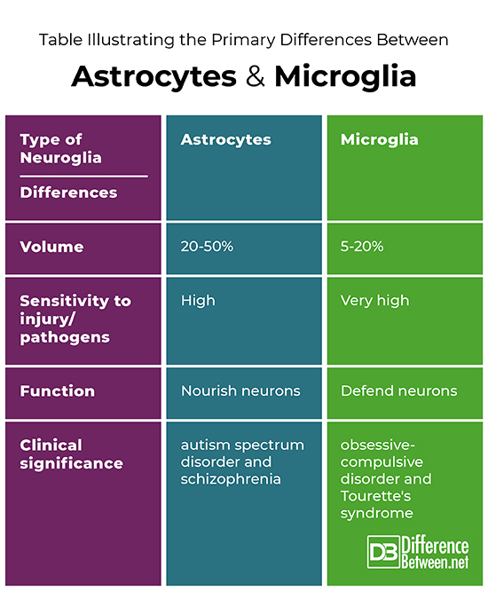
Although both are types of neuroglia, astrocytes are macroglia and more prevalent, making up 20-50% of the volume of the CNS as opposed to just 5-20% for microglia.
Both microglia and astrocytes respond to neuronal injury by proliferating, altering, mediating, and engulfing damaged or infected cells and subcellular elements. These changes represent the CNS tissue response to damage or degeneration. Microglia, however, have been shown to be more sensitive to pathogens.
Each type of neuroglia has distinct functions. So, although both offer physical and metabolic support to the CNS, astrocytes nourish neurons whereas microglia protect neurons from pathogens that have permeated the blood brain barrier, even if that defensive role results in collateral damage.
Each type of neuroglia also has distinct psychiatric significance. Whereas astrocytes have been shown to be involved in neurodevelopment disorders such as autism spectrum disorder and schizophrenia, microglia have been shown to be involved in the pathophysiology of obsessive-compulsive disorders and Tourette’s syndrome.
Table illustrating the primary differences between astrocytes and microglia

Summary
Astrocytes and microglia are two different types of neuroglia that support the CNS. While the more prolific astrocytes nourish cells in the CNS, including other neuroglia, microglia protect and defend neurons from pathogens that have permeated the blood brain barrier. Both assist with the repair of damaged neural tissue. Dysfunction of each neuroglia is associated with distinct psychiatric disorders, astrocytes with autism spectrum disorder and schizophrenia and microglia with obsessive-compulsive disorder and Tourette’s syndrome.
FAQ
Are microglia a type of astrocyte?
Microglia and astrocytes are distinct types of neuroglial cells distributed throughout the CNS and have a different genesis and function. Tian et al. notes that astrocytes are macroglia that modulate the entry of immune cells into the CNS via the blood–brain barrier and perform an executive-coordinating function in relationship to other neuroglia like microglia.
How do astrocytes interact with microglia?
Interaction between activated microglia and astrocytes plays a critical role in reducing neuroinflammation. Via oligodendrocytes, astrocytes activate microglia when unable to prevent pathogens from permeating the blood brain barrier. In turn, microglia activate and secrete molecular signals to trigger reactive astrocytes to reduce neuroinflammation.
What are oligodendrocytes and how are they different astrocytes and microglia?
Oligodendrocytes are another type of neuroglial cell that wrap a myelin sheath made of lipid and protein around axons, or nerve fibers, to support, insulate and facilitate the transmission of neural signals. So, unlike astrocytes that nourish neurons and microglia that protect neurons from pathogens, oligodendrocytes facilitate communication between neurons and glia, including microglia.
- Difference Between Ecchymosis and Erythema - August 15, 2022
- Difference Between Autobiographical Memory and Episodic Memory - August 1, 2022
- Difference Between Biological Drive and Social Motive - July 30, 2022
Search DifferenceBetween.net :
Leave a Response
References :
[0]Barker, A. J. and E. M. Ullian. "New roles for astrocytes in developing synaptic circuits." Communicative & Integrative Biology, vol. 1, no. 2, 2008, pp. 207–11. doi:10.4161/cib.1.2.7284.
[1]Cserép C., et al. "Microglia monitor and protect neuronal function through specialized somatic purinergic junctions." Science, vol. 367, no. 6477, January 2020, pp. 528–537. doi:10.1126/science.aax6752.
[2]Kreutzberg G. W. "Microglia, the first line of defence in brain pathologies." Arzneimittelforschung, vol. 45, no. 3A, March 1995, pp. 357–360.
[3]Swaminathan, N. "Brain-scan mystery solved." Scientific American Mind, vol. 7, no. 1. October 2008. doi:10.1038/scientificamericanmind1008-16.
[4]Tian, Li, et al. "Neuroimmune crosstalk in the central nervous system and its significance for neurological diseases." Journal of Neuroinflammation, vol. 9, July 2, 2012, p. 155. doi:10.1186/1742-2094-9-155.
