Difference Between Piecemeal Necrosis and Bridging Necrosis

Introduction
Necrosis refers to the loss or death of a cell, resulting in a cell which is no longer functional. Cell death is a normal process in the human body, but when it occurs in excess or in areas it should not then it becomes physically harmful. Necrosis is often found in acute and long-term disease of the liver. Given the persistent nature of liver disease, fibrosis progresses alongside necrosis among parts of the liver. The pattern in which necrosis presents itself provides medical professionals with important information about the nature of a patient’s liver.
Piecemeal necrosis (also known as interface hepatitis) and bridging necrosis (also referred to as confluent necrosis) are both types of necrosis occurring in the liver. Piecemeal necrosis is associated with a cell degenerating effect where bridging necrosis refers more to where in the liver cell loss is taking place. Associated with similar and different hepatic (liver) conditions, these types of necrosis are indicative of severe disease complications and can present acutely or over time.
This article will take a closer look into the differences between piecemeal and bridging necrosis.

Definition and Pathophysiology
Piecemeal Necrosis
Piecemeal necrosis is often referred to as interface hepatitis. This necrosis is associated with viral hepatitis, autoimmune hepatitis, and steatohepatitis. The physiology behind this necrosis relates to the degeneration or total loss of the limiting plate of liver cells. Ultimately, it involves inflammation processes with eroding effects and takes place at the lobular-portal-interface. This interface is the area where portal tracts and the hepatic parenchyma form a junction.
Bridging Necrosis
Bridging necrosis is also referred to as confluent necrosis. More specifically, this necrosis involves vascular structures. In terms of the liver, bridging necrosis involves the centrilobular veins (terminal hepatic venules) and the linkage to the liver portal tracts. An example of bridging necrosis can include when necrosis occurs across various zones in the lobes of the liver.
Potential Causes
Piecemeal necrosis is generally a feature of chronic viral hepatitis and long-term autoimmune hepatitis.
Bridging necrosis is caused mostly by a severe acute viral hepatitis, presenting most commonly in this condition. Bridging necrosis can also occur when the liver experiences injury, such as through alcohol, drugs, and other immediate injuring substances.
Clinical Presentation and Diagnosis
In biopsies (or samples) of liver cells, piecemeal necrosis shows a collected layer of hepatocytes around the portal tact. In long-term hepatitis patients, this is also shows and a following presentation is a state of fibrosis. When piecemeal necrosis takes place because of autoimmune associated hepatitis, then blood tests can show autoimmune antibodies too.
Piecemeal necrosis can affect parts of portal areas or the entire portal area.
Bridging necrosis is seen when cells are sampled (biopsied) and stained before placed under a microscope. When looking at the cells there will be a significant lack of collagen deposition and at a later stage there will even less original portal tract collagen present. Cell studies will also reveal that there is a collapsed reticulin framework in the zones which are necrotic and in the zones which have collapsed, there will be missing elastic fibres that should have been deposited.
Treatment
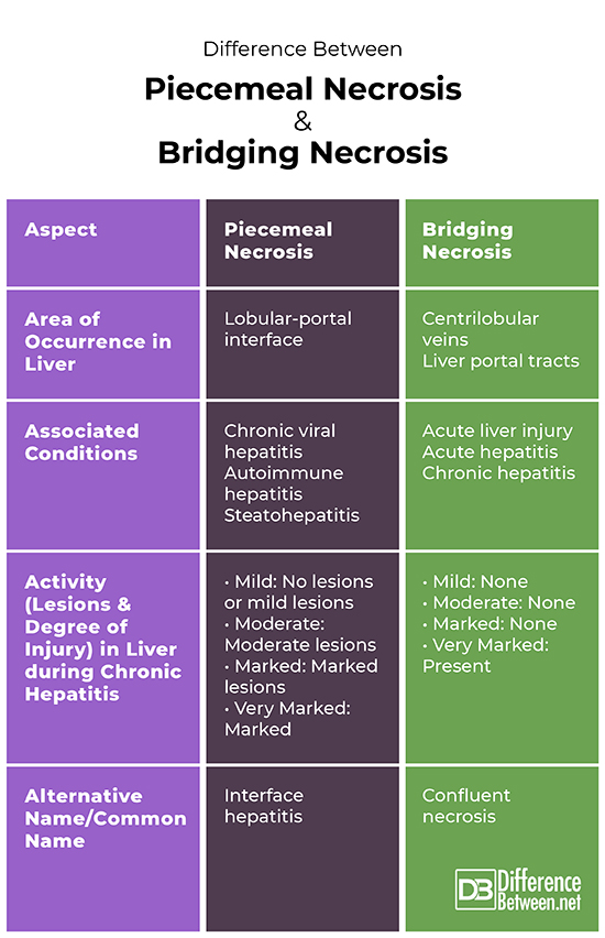
Table of comparison for piecemeal necrosis and bridging necrosis

Summary
Piecemeal necrosis and bridging necrosis are both manifestations of liver conditions. Where piecemeal necrosis is associated with chronic forms of hepatitis and autoimmune hepatitis, bridging necrosis is mostly associated with acute liver injuries but also occurs in long-term hepatitis.
Piecemeal necrosis occurs in the lobular-portal interface in the liver, where bridging necrosis occurs in vascular structures like centrilobular veins and liver portal tracts.
When diagnosing both types of necrosis, a sample of liver cells are investigated. Piecemeal necrosis shows that there is a collection of hepatocytes where bridging necrosis shows a lack of cell collagen and structural collapses.
Piecemeal necrosis is also referred to as interface hepatitis. Bridging necrosis is commonly referred to as confluent necrosis.
Looking at the degree of injury and lesions in the liver during chronic hepatitis, piecemeal necrosis doesn’t reflect generally in mild cases. In moderate cases, there is moderate piecemeal necrosis lesions or injury and in marked and very marked cases, there is a marked presence. Looking at bridging necrosis activity presence in mild, moderate and marked in chronic hepatitis, there is an absence. Very marked chronic hepatitis can expect a presence of lesions and a degree of injury associated with bridging necrosis.
FAQ
What is piecemeal necrosis?
Piecemeal necrosis is often referred to as interface hepatitis. This necrosis is associated with viral hepatitis, autoimmune hepatitis, and steatohepatitis. The physiology behind this necrosis relates to the degeneration or total loss of limiting plates (also known as hepatocytes). Ultimately, it involves inflammation processes with eroding effects and takes place at the lobular-portal-interface. This interface is the area where portal tracts and the hepatic parenchyma form a junction.
What are the different types of bridging necrosis?
Bridging necrosis involves more than one zone inside the liver lobes. It can occur in different types, these include central-central bridging necrosis and portal-portal bridging necrosis.
What does bridging necrosis mean?
Bridging necrosis refers to the loss of cells occurring in the liver. This type of necrosis occurs specifically in the portals, tracts or vascular structures within the liver and occurs across zones. The crossing over zones and structures is where the term “bridging” comes from, as just as a bridge on the roads, it suggests a reaching across two regions.
What causes piecemeal necrosis
Piecemeal necrosis can be caused when suffering with chronic viral hepatitis and long-term autoimmune types of hepatitis.
- Difference Between a Cochlear Implant and Normal Hearing - October 4, 2022
- Difference Between Obstructive and Restrictive Spirometry - September 11, 2022
- The Difference Between White Box and Black Box Testing - September 11, 2022
Search DifferenceBetween.net :
References :
[0]Human Path. “Bridging necrosis”. 2004. https://www.humpath.com/spip.php?article2349
[1]Kamal, G.I. “Pathologic Features of Chronic Hepatitis”. American Journal of Clinical Pathology, vol. 113, 2000,pp. 40-55.
[2]Krishna, Murli. “Patterns of necrosis in liver disease”. Clinical Liver Disease, vol. 10, no. 2, 2017, pp: 53-56.
